Call: 08071930543
 Send Inquiry
 Send Inquiry
Pathological Research Microscope
Pathological Research Microscope Specification
- Focus System
- Coaxial coarse and fine focusing
- Features
- Anti-fungal optics, ergonomic design, sturdy metal frame, wide-field observation, suitable for research and education
- Spare Parts
- Fuse, bulb, lens cleaning cloth, power cord
- View Head
- Trinocular, sliding type
- Theory
- Pathological research microscope designed for diverse clinical and laboratory applications.
- Drawtube
- Trinocular
- Sensor
- CMOS Sensor
- Resolution
- 1920 x 1080 pixels
- Interface
- USB 2.0, HDMI
- Frame Rate
- 30fps at full resolution
- Focal Distance
- 45 mm
- Magnification
- 40X to 1000X
- Dimensions
- 400 x 235 x 420 mm
- Focus Range
- 8 mm
- Eyepieces
- WF10x/20mm, high eye-point
- Eyepiece Tube
- Inclined 30, rotatable 360
- Illumination
- LED, 3W with adjustable brightness
- Coarse Adjustment Range
- 30 mm
- Fine Adjustment Range
- 0.002 mm
- Working Stage
- Double layer mechanical stage, 140 x 140 mm with 75 x 50 mm moving range
- Still Image Capture Resolution
- 2 Megapixel
- Video Capture Resolution
- Full HD 1080p
- Image Format
- JPG, BMP, TIFF
- Interpupillary Distance
- 55-75 mm adjustable
- Objective Achromatic
- 4x, 10x, 40x (S), 100x (S, Oil) DIN
- Condenser
- Abbe condenser N.A. 1.25 with iris diaphragm and filter
- Light Source
- Built-in LED, 100-240V AC
- Power Supply
- AC 100-240V/50-60Hz in-built adapter
- Ambient Temperature Range
- 10°C to 40°C
- Packing
- Supplied in thermocol box with instruction manual
- Color
- White and black
- Humidity Tolerance
- Up to 80% RH
- Body Material
- Heavy-duty die-cast aluminum
- Nosepiece
- Quadruple revolving nosepiece
- Stage Movement
- Low-position coaxial X-Y controls
Pathological Research Microscope Trade Information
- Main Domestic Market
- All India
About Pathological Research Microscope
Pathological Research Microscope
| Usage/Application | Laboratory |
| Brand | Royal Scientific Works |
| Eye Piece | 5X, 15X Huygenian and 10X WF |
| Objectives Achromatic | 4X, 10X, 40 or 45x and 100x Oil Immersion spring loaded |
| Lighting Type | Halogen Bulb |
| Bulb Voltage | 6 V |
Advanced Optical Precision
Equipped with a high-resolution CMOS sensor and Full HD capability, this microscope achieves exceptional clarity across multiple magnifications, from 40X to 1000X. The anti-fungal optics preserve lens quality while the Abbe condenser and wide-field eyepieces guarantee crisp imaging for pathology and research.
Ergonomic Design & Sturdy Construction
The heavy-duty die-cast aluminum body ensures durability, while the low-position coaxial controls and double-layer mechanical stage enhance user comfort during extensive observations. The entire system is protected with an anti-fungal coating, making it highly suited for varied environmental conditions.
User-Friendly Digital Integration
Designed with a trinocular drawtube and interfaces for USB 2.0 and HDMI, digital imaging and documentation are seamless. Whether capturing still images or recording Full HD videos at 30fps, the microscope offers robust, flexible connectivity for research and educational analysis.
FAQs of Pathological Research Microscope:
Q: How does the Pathological Research Microscope benefit pathological and clinical research?
A: With a magnification range of 40X to 1000X, Full HD digital imaging, and anti-fungal, wide-field optics, this microscope delivers superior sample visualization and accurate documentation, essential for reliable research and diagnostic processes.Q: What stage movement features make sample analysis easier?
A: Its low-position coaxial X-Y controls and double-layer mechanical stage (140 x 140 mm) with a 75 x 50 mm moving range enable smooth, precise sample navigationcrucial for detailed examination and sequential analysis.Q: When is digital documentation recommended during usage?
A: For training, research presentations, or archiving, use the trinocular drawtubes CMOS sensor and USB/HDMI interfaces to capture high-resolution still images (2MP) or Full HD videos directly onto compatible devices whenever visual records are needed.Q: Where can this microscope be installed and operated?
A: It is designed for laboratories, educational institutions, clinics, and research facilities. Thanks to its broad ambient operating temperature range (10C to 40C) and tolerance for up to 80% relative humidity, it adapts well to diverse environments.Q: What is the process for adjusting magnification and viewing angles?
A: Rotate the quadruple revolving nosepiece to select objective lenses (4x, 10x, 40x, 100x oil). Adjust the inclined, rotatable trinocular eyepiece tube to suit your comfort and use the coarse (30 mm) and fine (0.002 mm) focusing controls for sharp, clear imagery.Q: How does the built-in LED illumination enhance microscopy?
A: The 3W adjustable LED illumination ensures even, bright lighting with low heat output, improving sample visibility and reducing eye strain during extended observation sessions.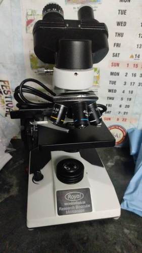
Tell us about your requirement

Price:
Quantity
Select Unit
- 50
- 100
- 200
- 250
- 500
- 1000+
Additional detail
Mobile number
Email

More Products in MICROSCOPES Category
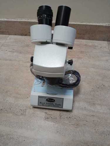
STEREO MICROSCOPE
Price Range 45000.00 INR / Unit
Minimum Order Quantity : 1 Unit
Theory : Other, Stereo microscopy enables observation of samples with threedimensional vision, suitable for biological and industrial inspection.
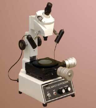
Tool Maker Microscope
Price Range 15000.00 - 30000.00 INR / Piece
Minimum Order Quantity : 1 , , Piece
Theory : Biological Microscope
Application : Lab Field
Material : Stainless Steel
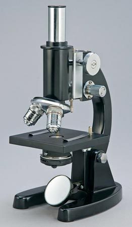
Student Microscope
Price 1660 INR / Piece
Minimum Order Quantity : 1 Piece
Theory : Biological Microscope
Application : Lab Field
Material : Stainless Steel
Dissecting Microscope
Price nan INR / Piece
Minimum Order Quantity : 1 Piece
Theory : Biological Microscope
Application : Lab Field
Material : Stainless Steel
"We are accepting only bulk quantity orders."
Contact Details
ROYAL SCIENTIFIC WORKS
GST : 06BOHPS6662C1ZS
- Village Keshopur, P.O Sambhalkha, Distt Ambala, Ambala Cantt - 133101, Haryana, India
- Phone :08071930543
Developed and Managed by Infocom Network Private Limited.
